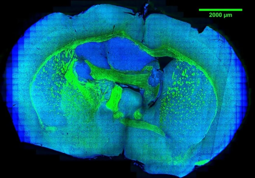
Tumor metabolism supports the abnormal survival and growth of malignant cells by providing energy, biomolecular precursors, and reducing equivalents. It is regulated by a combination of cell-intrinsic and cell-extrinsic factors. Upregulated syntheses of new biomolecules in cancer cells are the hallmark biological characteristic in tumor. However, current techniques such as MRI and PET have limited spatial resolution to observe caner metabolism and its heterogeneity at subcellular resolution. To demystify the new synthesized biomolecules’ spatial distribution inside tumor, we applied bioorthogonal chemical imaging to visualize newly synthesized biomolecules in tumors after in vivo delivery of alkyne or isotope labeled metabolites into tumor-bearing mice. This method opens a door to study tumor metabolic heterogeneity and to observe metabolic competition or cooperativity among stromal, immune, and tumor cells with subcellular resolution.
L. Zhang and W. Min. Bioorthogonal chemical imaging of metabolic changes during epithelial–mesenchymal transition of cancer cells by stimulated Raman scattering microscopy. J. Biomed. Opt. 22, 106010 (2017).
F. Hu, Z. Chen, L. Zhang, Y. Shen, L. Wei and W. Min. “Vibrational imaging of glucose uptake activity in live cells and tissues by stimulated Raman scattering”, Angew. Chem. Int. Ed. 54, 9821 (2015).