Glucose is a ubiquitous energy source for most living organisms. Its uptake activity closely reflects cellular metabolic demand in various physiopathological conditions. Extensive efforts have been made to specifically image glucose uptake, such as with positron emission tomography, magnetic resonance imaging, and fluorescence microscopy, but all have limitations. To visualize glucose uptake activity in live cells and tissues, we are performing SRS imaging on a novel glucose analogue labeled with a small alkyne moiety. Cancer cells with differing metabolic activities can be distinguished. Heterogeneous uptake patterns are observed with clear cell–cell variations in tumor xenograft tissues, neuronal culture, and mouse brain tissues. By offering the distinct advantage of optical resolution but without the undesirable influence of fluorophores, this method will facilitate the study of energy demands of living systems with subcellular resolution.
R. Long, L. Zhang, L. Shi, Y. Shen, F. Hu, C. Zeng and W. Min. Two-color Vibrational Imaging of Glucose Metabolism by Stimulated Raman Scattering. Chem. Commun. 54, 152 (2018).
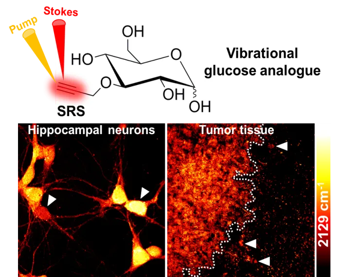
F. Hu, Z. Chen, L. Zhang, Y. Shen, L. Wei and W. Min. “Vibrational imaging of glucose uptake activity in live cells and tissues by stimulated Raman scattering”, Angew. Chem. Int. Ed. 54, 9821 (2015).