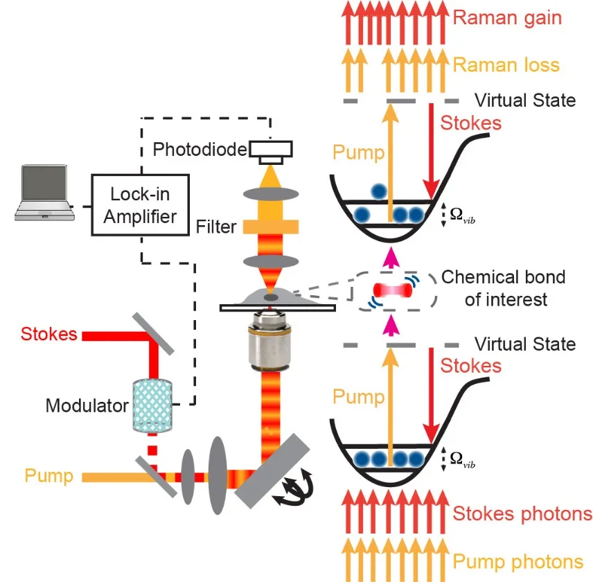
Chemical bonds are inherent targets for optical spectroscopy, offering power spectroscopic contrast for chemical imaging. Stimulated Raman scattering (SRS) microscopy has emerged as a highly sensitive and specific vibrational imaging technique. When the energy gap between two lasers (pump beam and Stokes beam) is resonant with the vibrational level of targeted chemical bonds, the joint action of the pump and Stokes fields stimulates (i.e., accelerates) the otherwise slow vibrational transition by 108 times. Spectroscopically, the SRS spectrum is identical to that of spontaneous Raman without the complication of nonresonant background, thus offering straightforward and robust spectral interpretations. The detection sensitivity of SRS is several orders of magnitude more sensitive than the conventional Raman microscopy. As a result, video rate SRS imaging is feasible. In addition, SRS signals are linearly dependent on analyte concentrations, allowing for quantitative analysis. Moreover, the nonlinear nature and the adoption of near-infrared laser wavelengths allow SRS three-dimensional optical sectioning into deep tissues. After raster-scanning across the sample, one can produce a three-dimensional concentration map of the targeted chemical bonds in biological samples.
C. W. Freudiger*, W. Min*, B. G. Saar, S. Lu, G. R. Holtom, C. He, J. C. Tsai, J. X. Kang and X. S. Xie. “Label-free biomedical imaging with high sensitivity by stimulated Raman scattering microscopy”, Science, 322, 1857, (2008).
W. Min*, S. Lu*, S. Chong, R. Rahul, G. H. Holtom and X. S. Xie. “Imaging chromophores with undetectable fluorescence by simulated emission microscopy”, Nature, 461, 1105, (2009).
W. Min, C. W. Freudiger, S. Lu and X. S. Xie. "Coherent nonlinear optical imaging: Beyond fluorescence microscopy", Annu. Rev. Phys. Chem., 62, 506, (2011).